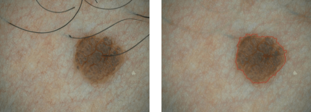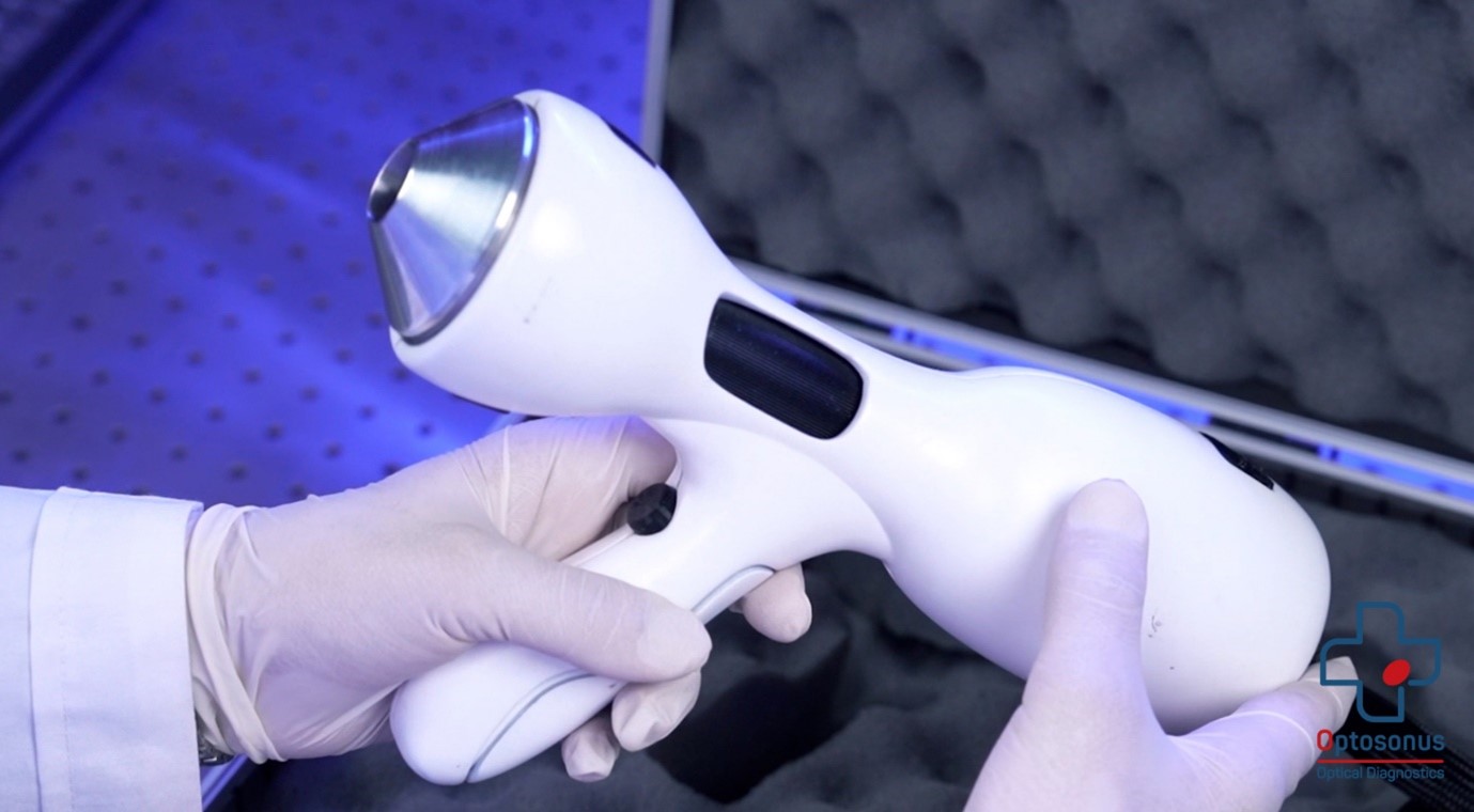
Dermatoscopy has a prominent position for skin examination, especially for pigmented skin lesions. Dermatologists can apply dermoscopes to see the color distribution and structures of lesions in epidermis. A dermoscope comprises a magnifying optical system and a light source illuminating the skin to be examined. The dermoscope is placed on the skin wherein the magnifying optical system helps to better see the skin lesion via an eye or video capturing system based on ccd image sensors. Using polarized illumination reduces the specular reflection so the operator does not require any refractive index matching oils or liquid that enhances the image quality, and it also makes the procedure of dermatoscopy easier. Optosonus invented a new polarized dermatoscope, OptoSkin, for imaging of epidermis and superficial dermis (increasing the optical depth of skin imaging). The target underlying this production is to design an optical system comprising adjustable cross-polarized illumination and magnifying parts which enables an operator to optimally utilize the dermoscope without great efforts to many regions of interest (ROI) on the skin, simultaneously.
Optoskin applies ABCD rules to score the skin lesion via measuring the diameter, area, and the colors of the lesion. Dermatologists can use ABCD score to find the potential skin cancer.